EPM – Part I: What is EPM and How Did My Horse Get It?
What exactly is EPM?
Equine protozoal myeloencephalitis, simply known as EPM, attacks the horse’s central nervous system and causes inflammation and damage to the brain and/or spinal cord. EPM in horses is often a confusing and hard-to-diagnose disease that has been scrutinized by equine researchers since the 1970s
Researchers have identified two different types of protozoal microorganisms as the cause of EPM. Sarcocystis neurona is the most common culprit. Neospora hughesi is less likely to be found, but when present causes just as much trouble as its fellow protozoan. It is entirely possible for both microorganisms to be present at the same time.
EPM is defined as a progressive, degenerative disease, which means as time passes, the inflammation can become widespread and the damage can increase in severity. Once affected, the function of the tissues in the central nervous system may continue to deteriorate. EPM in horses can be a fatal disease.
EPM is one of the most commonly diagnosed neurological diseases in horses. Interestingly, it has not been reported in mules, donkeys, or other non-horse equids. EPM has been found in horses from 2 months to 24 years of age. It is most often diagnosed in horses between 1 and 6 years old. It can be seen at any time of the year. The protozoa that cause the disease are widespread and horses may be continuously exposed during their lifetimes. It has been suggested that 50% of the equine population in the USA has been exposed. The good news is, less than 1% of the horses exposed to these disease-carrying protozoa develop clinical EPM.
How or why some horses are able to fight off the invading protozoa is unknown. Are some horses able to develop immunity? Do clinical signs always appear directly after exposure? Can the microorganisms lie dormant until an opportune moment arises? These are the questions researchers are asking. It has been proposed that there is a combination of factors involved. There may be a variation in the potency of the invading protozoa and/or the ability of the horse’s immune system to mount a defense. There is some evidence that previously infected horses might harbor the microorganism within the central nervous system and that stress can lead to the development of clinical symptoms. Hopefully ongoing research will answer these questions and unravel some of the mystery surrounding this disease.
The role hosts play in spreading EPM in horses
To get a feel for how EPM is spread it is important to understand the role multiple hosts play in the life cycle of the causative agent. The causative agent is a pathogenic microorganism that is capable of causing disease. In this case there are two causative agents, Sarcocystis neurona and Neospora hughesi. Recent research has revealed that both protozoa have a wide distribution so both need to be considered. Since Sarcocystis neurona is the most common agent, we know the most about its life cycle.
The life cycle of Sarcocystis neurona
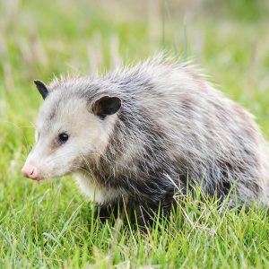
A host is the animal or plant on or in which a microorganism lives. There are several types of hosts involved in spreading EPM. The primary host is the host in which the microorganism reaches a mature state and sexually reproduces. The opossum is considered the primary host in spreading EPM. A secondary host provides a safe harbor for a period of time, usually while some developmental stage is completed. There are many secondary hosts involved in the life cycle of Sarcocystis neurona, including cats, armadillos, skunks, raccoons, and sea otters. The primary and secondary hosts are not attacked by the protozoa so they don’t get ill. A dead-end host is one that ingests the microorganism and then gets sick. Dead-end hosts do not pass the microorganism on to others. The dead-end host in this case is the horse.
As the primary host we will start with the opossum. They harbor an environmentally resistant stage of the protozoa in their digestive tract that passes out in their feces. The secondary host eats the opossum’s feces and ingests the protozoa. The protozoa then develop in the body of the secondary host until they reach a resting stage in the muscle tissue. After the secondary host dies, the opossum comes along and consumes the carrion along with the contaminated muscle tissue. Once back in the opossum’s digestive tract, the protozoa mature and produce sporulated oocysts (fertilized cells), which are excreted in the feces. The feces containing these oocysts contaminate a horse’s feed or water, and the horse ingests them. The oocysts migrate out of the equine digestive tract by hitching a ride on the horse’s white blood cells, where they enter the CNS via the blood-brain barrier. The protozoa then replicate within the neuronal cells, causing inflammation and cell damage.
Researchers are still learning about the life cycle of Neospora hughesi and have yet to identify the definitive host.
In part II of our series, we will review the symptoms of EPM in horses and what test your veterinarian might use to make an accurate diagnosis.


17 Comments
In you comment “Interestingly, it has not been reported in mules, donkeys, or other non-horse equids.” I would like to say that Mules can get EPM. My mule has been blood tested and diagnosed is positive. They carries the same symptions as the horse.
Hi Julie,
Thanks for the information. There has been very little work done on EPM in donkeys and mules so it isn’t out of the realm of possibility that your mule could have tested positive and presented with the clinical disease.
Upon completing a review of the most recent literature, it seems it is still generally accepted that outside of horses, other non-horse equids are not susceptible to the clinical disease. Exposure does not always equate to clinical symptoms, and other diseases and vitamin deficiencies can mimic clinical symptoms, which make EPM a challenge to diagnose.
It is interesting to note that per the AAEP’s most recent guidance, testing of the cerebrospinal fluid (CFS) and serum is recommended for a definitive diagnosis.
References:
Mendoza FJ, Toribio RE, Perez-Ecija A. Donkey Internal Medicine—Part II: Cardiovascular, Respiratory, Neurologic, Urinary, Ophthalmic, Dermatology, and Musculoskeletal Disorders. In J Equine Vet Sci. 2018 Jun; 65: 86–97.
Dubey JP, Lindsay DS, Saville WJ, Reed SM, Granstrom DE, Speer CA. A review of Sarcocystis neurona and equine protozoal myeloencephalitis (EPM). In Vet Parasitol. 2001:95:89-131.
AAEP Infectious Disease Guidelines: Equine Protozoal Myeloencephalitis
https://aaep.org/sites/default/files/Guidelines/EPM_Disease_Guidelines_2021.pdf
Our 21 year old mule has also just positive for EPM. He started showing symptoms, tripping.
Tested for EPM, LYMES and VIT E DEFICIENCY. Lyme and Vit E were both negative. EPM titer was high.
Our two mini donkeys ages 8 and 12 were tested quantitatively blood positive. Only the 12 yo has developed symptons. (Severe swaying, stumbling, falling). Three rounds of Marquis seems to have at least slowed the progression. Im told the brain and cns damage is likely irreversible. Its been a terrifying time. Any further info is appreciated.
Your treating veterinarian will be able to give you the best prognosis. Every case of EPM is different depending on the severity and location of the lesions, and how quickly treatment was started. Some horses with severe symptoms recover normal muscle and nerve function, while others remain impaired.
I understand the tests for epm aren’t always accurate. Are certain types better than others? Is there research as to which drug is the best to treat?
Dear Janet,
I suggest you read part II of this article. I think you will find it helpful. https://kppusa.com/2018/05/09/epm-symptoms-diagnosis-and-treatment/
Testing both the cerebrospinal fluid (CSF) along with a blood serum sample is the gold standard in diagnosis. CSF is collected via a spinal tap, which can be risky and expensive so it isn’t done very often. In most cases, a positive serum IgG test combined with signs of neurological deficits and a history consistent with exposure to EPM will serve as a positive diagnosis.
There are several drugs available to treat EPM. Marquis (ponazuril) and Protazil (diclazuril) have both been approved by the FDA, so they are research-proven treatments. ReBalance (sulfadiazine and pyrimethamine) has been tested by several researchers and is thought to be effective in some cases.
Your veterinarian will work with you to pick the best medication for your horse’s situation. Sometimes multiple treatments are recommended over a period of time, depending on how your horse is recovering. The earlier you can start treatment, the better. Severely damaged nerve tissues may not recover.
Along with whatever drug your vet prescribes, supportive therapy with natural vitamin E is also recommended. Talk to your vet about Elevate® W.S., liquid natural vitamin E.
Elevate W.S. is the only vitamin E research-proven to cross the blood-brain barrier.
• Passes through the blood-brain barrier to impact CSF
• Can be fed at high levels for long periods of time with no adverse effects
• Builds immune response
• Supports healing of damaged neurological tissues
I hope this answers your question.
Best regards,
Karen at KPP
So if my horse is positive, should I get rid of all my hay and start fresh? Can the hay be tested to see if it is contaminated? The hay is stored in possum proof buildings, but I cannot vouch for the storage before I buy it, from a dealer who ships it in from out west (Montana/Utah/etc.)
Horses can be exposed to EPM in many ways. Contaminated pasture grass, concentrates, hay or water sources are the typical culprits. The key is to make your farm as unfriendly to possums as possible!
Possums are attracted to barns that contain readily available food, such as leftover feed in pastures, cat or dog food that is left out, human garbage, or open bins of feed. They often use hay as a nesting site in barns where food is available. Unless the possums have been living in your barn and defecating on your hay, it is unlikely to be the source of exposure.
Make sure you clean up any feed or hay your horses leave behind if you feed outside. Check and clean your waterers daily and change the water in stock tanks often. Don’t leave any human garbage or pet food out overnight. Restrict horses from natural water sources that might become contaminated. Remove animal carcasses on your property (or on the road near your property) and dispose of them. The less the possums roam around your farm the lower your horses’ exposure to their feces.
I realize this reply is dated, but Montana doesn’t have opossums. You wouldn’t need to worry about contamination coming from here as long as your hay doesn’t make any stops in States where they might crawl on the trailer. Utah does have them though, FYI.
Hi Janet,
You are correct that the opossum’s range does not include many of the western states. However, hay can be contaminated once it arrives and is stored on the horse farm in areas where opossums reside, so even if a horse owner is shipping in western hay, it could still become contaminated while it is being stored on the farm.
It actually HAS been identified in donkeys.
Yes, a researcher in Brazil found serological evidence that Donkey’s have been exposed to EMP, but it is our understanding that all these Donkey’s were asymptomatic. I don’t think there is a definitive answer as to how the disease presents in the Donkey.
Prevalence of antibodies against Neospora spp. and Sarcocystis neurona in donkeys from northeastern Brazil
Abstract
Sarcocystis neurona and Neospora hughesi are coccidian protozoa that can cause neurological illness in horses in America. In this study we report seroprevalence of Neospora spp. andS. neurona in sera of 333 donkeys from the northeastern region of Brazil. Antibodies to Neospora spp. were detected in 2% (7 donkeys) of 333 sera tested by the indirect fluorescent antibody test (IFAT) with a cut-off dilution of 1:40. Antibodies to S. neurona were found in 3% (10 donkeys) of the samples tested by IFAT (cut-off ≥50) and 21% (69 donkeys) by the direct agglutination test (SAT ≥50). The SAT and IFAT results for S. neurona showed a poor concordance (value of Kappa=0.051). This is the first report ofNeospora spp. antibodies in Brazilian donkeys and the first detection of antibodies against S. neurona in this animal species.
Full paper: http://www.scielo.br/scielo.php?script=sci_arttext&pid=S1984-2961201600010010
Once you horse has EMP is it treated everyday, weekly, monthly or yearly? Does treatment make it disappear or keep it dorment , or are there flareup. Thank you Kelley Simpson
Hello Kelley,
Treatment protocols will vary in frequency and length of time with individual cases. The severity of symptoms, drug of choice, and the horse’s response to treatment are all taken into consideration. We recommend you work closely with your veterinarian when determining a treatment plan for your horse.
It is my understanding that once a horse is exposed to EPM, it persists in the body. Some horses never show symptoms and others do. Some horses have one episode, recover, and never have symptoms again; others have periodic flareups, and some never recover fully. Recovery depends on the extent of the initial damage caused by the disease. It is thought that flareups may be related to the ability of the horse’s immune system to keep the protozoa in check.
Best regards,
Karen
Pingback
[…] spinal cord. EPM is passed on to the horse when they consume feed contaminated by opossum feces. (See: EPM – Part I: What is EPM and how did my horse get it?) EPM is defined as a progressive, degenerative disease, which means as time passes, the inflammation […]
Pingback
[…] EPM – Part I: What is EPM and How Did My Horse Get It? […]